



Elucidating the mechanism by which protein glycan deficiency leads to disease – Exploring the causes of disease from gene variants in undiagnosed diseases – (Glycan & Life System Integration Center Director Professor Shoko Nishihara)
[Key points of the announcement]
- While genetic variant information is accumulating rapidly, the link between many of these variants and diseases remains unclear.
- We have identified cases where gene variants can lead to the loss of protein "glycans," impairing their function and potentially causing disease.
- Studies have shown that loss of sugar chains by the dephosphorylation enzyme ACP3 can impair enzyme function, protein stability, and extracellular secretion, potentially leading to impaired nerve development.
【overview】
The research group led by Professor Noriyoshi Manabe and Director Yoshiki Yamaguchi of the Institute of Molecular Biomembranes at Tohoku Medical and Pharmaceutical University, together with Director Shoko Nishihara of Soka University Glycan & Life System Integration Center, Director Masato Kaname of the Genome Medical Research Division at the National Center for Child Health and Development, Professor Junichi Furukawa of the Core Research Institute for Glycos, Nagoya University, and Professor Yoshihiro Akimoto of Kyorin University, are conducting research to analyze genetic data from patients with diseases of unknown cause (undiagnosed disease) and investigate the causes of these diseases.
This time, from over six million genome changes (variant 1) data on undiagnosed diseases collected by the National Center for Child Health and Development, we identified variants of genes that affect the 'glycans,' markers of proteins. Furthermore, we focused on the enzyme ACP3, which helps remove phosphate in the body, and conducted a detailed investigation. It was revealed that when glycans are lost due to this variant, their function as enzymes, stability as proteins, and secretion outside cells are impaired, potentially leading to disease.
These research results were published on October 16, 2026, in the scientific journal iScience, published by the American academic publisher Cell Press. This research was supported by AMED (Japan Agency for Medical Research and Development) Practical Research on Intractable Diseases (Project Management Numbers 22ek0109446, 24ek0109760, 25ek0109815, 26ek0109886) and Grant-in-Aid for Scientific Research (26K02160).
[Research background]
In recent years, advances in gene analysis technology have led to the accumulation of a vast amount of information on gene variants (Figure 1). However, less than half of these variants are clearly understood as either "causing disease" or "having no effect," and the significance of more than half of the gene variants remains unknown.
Among gene variants, there are those called "missense mutations 2" that alter the amino acids that make up proteins. These are known to affect the three-dimensional structure of proteins and the "glycans" that are attached to them as markers. If we can pinpoint that variants of unknown significance are the cause of disease, it may lead to the development of new treatments.
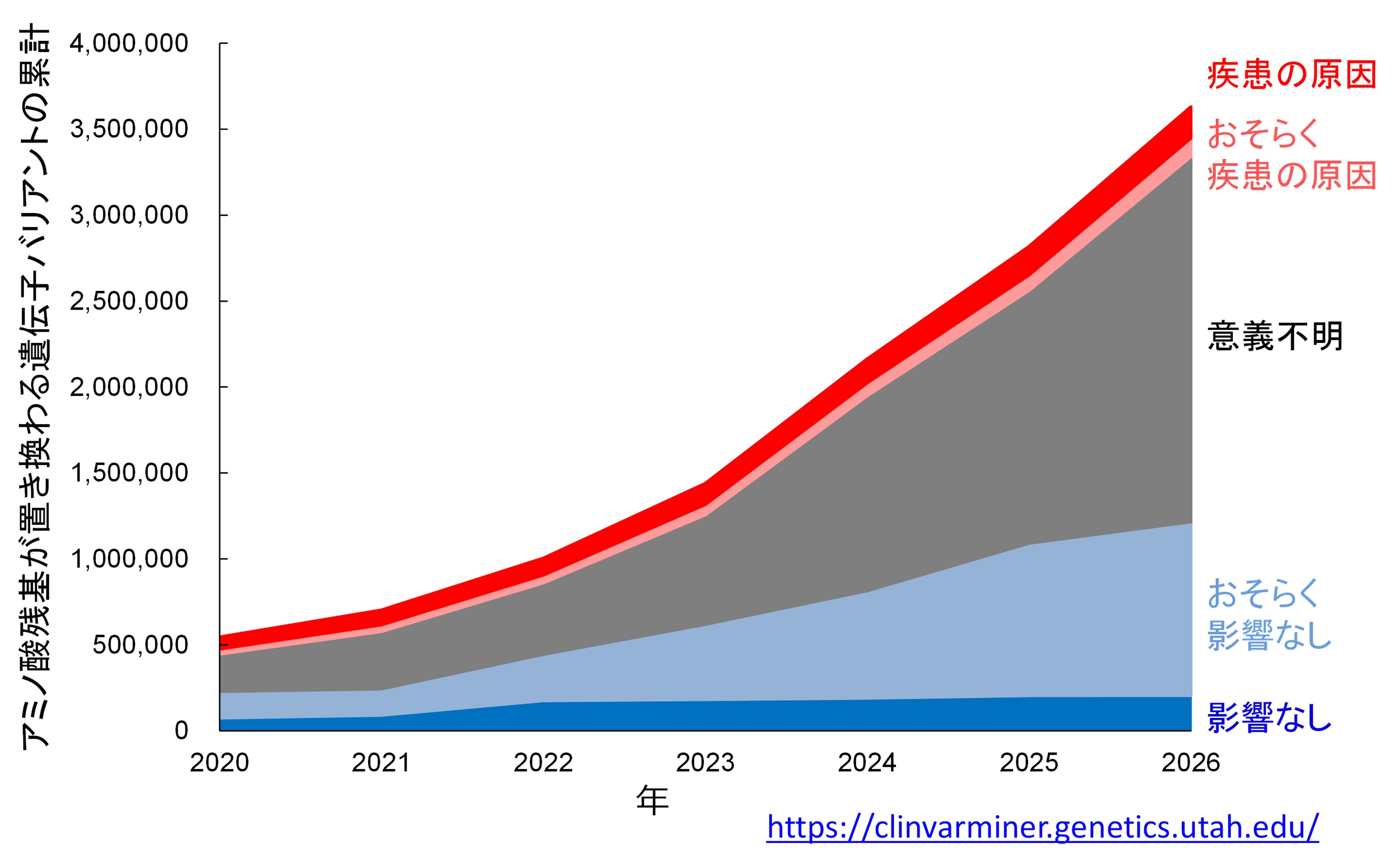
Genetic variants can be categorized into those found in healthy individuals with no effect, and those that cause disease. However, many reported variants are classified as "of unknown significance" because they cannot be categorized into either category. Evaluating the relationship between each of these variants and disease is crucial for accurate diagnosis, better understanding of diseases, and the development of treatments.
[Research content]
First, we narrowed down the missense mutations (variants) that are thought to cause abnormalities in glycosylation from over 6 million variant data points for undiagnosed diseases accumulated at the National Center for Child Health and Development. From these, we focused on three patients who had a variant (T335M) in which the 335th amino acid (threonine) of the enzyme ACP3, which removes phosphate, was replaced with methionine. When this variant occurs, the glycan that should normally be attached to the asparagine at the 333rd position of ACP3 is lost. Since all three patients showed abnormalities in neurodevelopment, we hypothesized that this glycan deficiency might be the cause of their disease and proceeded with a detailed analysis.
Therefore, we prepared recombinant proteins 3 produced by cells, including normal (wild-type) ACP3 and ACP3 modified to lack glycans, and compared them from various angles, such as enzyme function, location within the cell, protein degradation resistance (protease resistance test 4), and glycan structure analysis using mass spectrometry 5.
As a result, we confirmed that the T335M variant common to the patients lacked the glycosylation seen in the wild type. ACP3 with this variant was hardly secreted outside the cell (into the culture medium) and remained inside the cell. Further examination revealed that it could not reach the Golgi apparatus 6, the pathway for protein ejection from the cell, but remained in the endoplasmic reticulum 7, which is just before it. Furthermore, this protein that remained inside the cell had almost lost its original enzymatic activity. Experiments to examine its resistance to degradation also showed that it was more fragile and had an unstable three-dimensional structure compared to the wild type.
Furthermore, analysis using mass spectrometry revealed that the only type of glycan lost in this variant is a type called "high-mannose glycan 8". On the other hand, at the sites where other glycans are attached to ACP3, another type of glycan called "complex glycan 9" was predominant.
Furthermore, we used a computer simulation technique called molecular dynamics simulation10 to analyze the effect of glycans on the three-dimensional structure of ACP3. As a result, we found that the high-mannose glycan attached to asparagine at position 333 forms a stable network of bonds (hydrogen bonds) with the surrounding amino acids, acting as a nearly immobile "support." This result indicates that the glycan of Asn333 acts as a "structural clamp" that supports the structure of ACP3.
These results reveal that the T335M variant of ACP3, commonly observed in patients, exhibits reduced structural stability due to abnormal glycosylation, leading to loss of enzymatic function and impaired extracellular secretion. In particular, it is suggested that high-mannose glycans play a crucial role in maintaining the structural stability of ACP3. These molecular-level changes are thought to be involved in the development of neurological symptoms observed in patients (Figure 2).
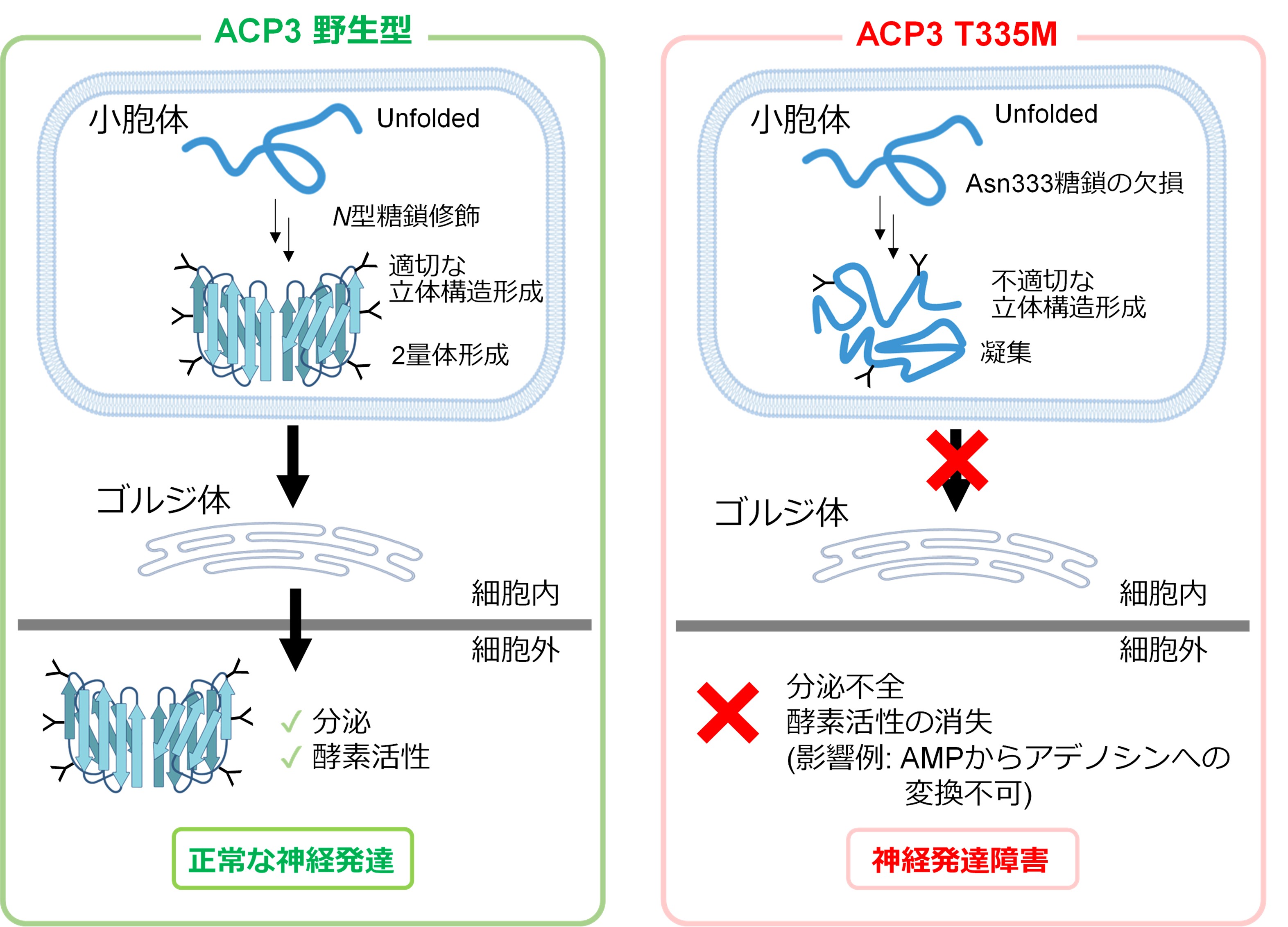
The T335M variant resulted in the loss of glycosylation at asparagine position 333, causing ACP3 to remain in the endoplasmic reticulum and aggregate, leading to structural instability and potentially losing its function as an enzyme that removes phosphate groups.
[Future Outlook]
This study focused on patients with the ACP3 T335M variant, but we have identified over 1,000 other variants that may be caused by abnormalities in modified glycans. We believe that clarifying the relationship between each of these variants and the disease will lead to the proposal of future treatments, so we will continue our analysis.
[Paper Title]
- Title: Loss of the Asn333 high-mannose N-glycan impairs ACP3 folding and drives disease-dysfunction associated
- (Japanese name): Loss of high-mannose N-linked glycans in ACP3 inhibits proper three-dimensional structure formation and causes functional impairment leading to disease.
- Publication: iScience
- Authors: Noriyoshi Manabe 1, Kazuyoshi Itoh 2, Shiho Ohno 1, Ayumi Miura 1, Aika Miyata 1, Sora Yashima 1, Sachiko Komatsu 3, Tsutomu Fujimura 3, Hisatoshi Hanamatsu 4, Jun-ichi Furukawa 4, Kiyohiko Angata 2, Akira Togayachi 2, Kiyoko F. Aoki-Kinoshita 2,4, Yoshihiro Akimoto 5, Tadashi Kaname 6, Shoko Nishihara 2, and Yoshiki Yamaguchi 1,
- Author affiliations: 1 Tohoku Medical and Pharmaceutical University - Molecular Biomembrane Research Institute, 2 Soka University -Glycan & Life System Integration Center, 3 Tohoku Medical and Pharmaceutical University - Faculty of Pharmacy, 4 Nagoya University - Glyco-Life Core Research Institute, 5 Kyorin University - School of Medicine, 6 National Center for Child Health and Development
[Explanation of Terms]
1. Variant: An individual difference or change in the DNA sequence. Some variants can cause disease, while others have no effect on the body.
2. Misssense mutation: A mutation in which one of the amino acids that make up a protein is replaced by another amino acid due to a change in the gene.
3. Recombinant proteins: Proteins that are artificially obtained by manipulating genes to make cells produce a target protein.
4. Protease Resistance Test: This experiment evaluates the stability of a protein's three-dimensional structure by examining its resistance to degradation by proteases, enzymes that break down proteins.
5. Mass Spectrometer: A device that precisely measures the mass of molecules. It is used to study the structure of proteins and glycans.
6. Golgi apparatus: An organelle within a cell that processes sugar chains attached to proteins and prepares them for release outside the cell.
7. Endoplasmic reticulum: An organelle within the cell where proteins are synthesized and folded into their correct three-dimensional structure. Proteins that do not fold properly remain here.
8. High-mannose glycans: These are relatively simple glycans consisting of many mannose sugar molecules linked together. They are found in the early stages of protein synthesis.
9. Complex Glycans: Glycans with more complex structures, formed when high-mannose glycans are processed in the Golgi apparatus.
10. Molecular Dynamics Simulation: An analytical method that calculates the movement of atoms and molecules on a computer based on physical laws, and reproduces changes in the three-dimensional structure of proteins and other molecules.
Faculty Information

Professor
Nishihara Shoko
Nishihara Shoko
- Specialized Field
Functional biochemistry, cell biology, developmental biology, general medical chemistry, glycobiology, stem cell biology, biochemistry, molecular biology
- Research theme
We are conducting research with the aim of clarifying the role of glycans in living organisms. We use various genetic engineering techniques to regulate the expression of glycan-related genes in Drosophila individuals, ES cells, iPS cells, cancer cells, cancer stem cells, human model cells, and organoids to clarify glycan functions. We also create knockout mice for some genes and conduct analysis.
(1) Analysis of glycan-related genes in Drosophila; elucidation of physiological functions of glycans conserved across species
Drosophila is the most advanced model animal in genetics. We focus on the "physiological activity of glycans conserved across biological species" and elucidate the role of glycans in the development of organisms through phenotypic analysis of mutants and knockdowns of glycan-related genes in Drosophila, as well as biochemical and molecular biological analysis. In particular, we are currently analyzing glycosyltransferases required for the maintenance and differentiation of blood stem cells, and glycan structures required for the formation of nerve axons.
(2) Elucidation of glycan functions in human and mammalian pluripotent stem cells (ES cells, iPS cells) and organoids
(1) We will use cultured cells, mainly embryonic stem cells, to examine whether the glycan functions revealed in the previous study are common to humans and other mammals. Specifically, we aim to "elucidate the role of glycans in stem cell maintenance and differentiation" by targeting ES cells and iPS cells. In this project, we first revealed in 2008 that glycans (heparan sulfate) are involved in the maintenance of ES cells. We have further developed this to reveal that the four glycan structures mentioned above are necessary for maintaining the naive pluripotent state. We are currently expanding the scope of our analysis to include various other glycans. There are still not many examples of functional analysis of glycans in embryonic stem cells, and we are playing a pioneering role in this field.(3) Functional analysis of PAPS transporter knockout mice
PAPS transporters are essential for the sulfation of glycans and proteins, and without them, each molecule cannot be modified by sulfation. We first isolated and identified them in 2003. Currently, we are creating knockout mice and analyzing them. We have found that these mice develop various diseases, and we are currently analyzing the mechanisms of their onset.
(4) Functional analysis of glycan-related genes involved in undiagnosed diseases
Based on the analysis conducted so far, it has been predicted that glycans are involved in many rare undiagnosed diseases, so we have begun analyzing these as well. We will analyze the loss of function of glycan-related genes with mutations that have been found to be associated with diseases using model organisms, stem cell differentiation systems, organoids, etc., to clarify the relationship with diseases.